
Laser-based refractive procedures are a group of ophthalmic techniques that aim to change the way the eye focuses light, typically by reshaping the cornea or altering internal optics. These procedures use controlled laser energy or laser-assisted tools to adjust corneal curvature or tissue profile, with the intent of reducing dependence on corrective lenses. The underlying concept involves precise measurements of refractive error and corneal anatomy, followed by a targeted tissue modification performed under local anaesthesia. Outcomes can vary by technique, eye anatomy, and healing response, so the aim is typically to improve unaided visual acuity rather than to promise a specific result.
Practitioners select among several procedural approaches based on diagnostic measurements, corneal thickness, and patient preferences. Some approaches create a thin corneal flap before applying laser energy, while others work on the corneal surface without a flap. Different laser platforms and guidance systems—such as femtosecond and excimer sources, and wavefront- or topography-guided treatments—may be used to plan and execute corrections. Informed consent and pre-procedure assessment usually cover potential outcomes, risks, and the possibility that additional adjustments may be considered later.
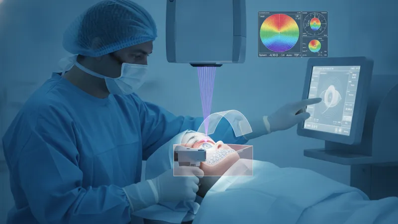
Comparisons among these procedures often focus on the structural approach to the cornea, wound healing patterns, and typical recovery timelines. For example, flap-based methods can allow faster visual recovery in many patients, while surface ablation methods may avoid flap-related considerations but often have a longer initial recovery period. Lenticule extraction aims to reduce surface disruption but requires specific laser platforms. When comparing techniques, clinicians typically evaluate corneal thickness, the degree and stability of refractive error, occupational or lifestyle factors, and diagnostic imaging results to identify plausible options rather than to assert a single preferred approach.
Laser and diagnostic technologies influence planning and potential outcomes. Instruments such as corneal topographers, tomographers, and aberrometers measure curvature, elevation, thickness, and higher-order aberrations that may affect visual quality. Treatment planning often integrates these data with manifest and cycloplegic refractions to create a customized ablation profile. Wavefront-guided and topography-guided protocols may aim to address pre-existing optical irregularities, and different laser platforms vary in pulse frequency, spot size, and delivery patterns, which can affect tissue interaction and procedural timelines.
Eligibility considerations commonly include stable refractive error over time, adequate corneal thickness for the intended correction, absence of progressive corneal disease, and reasonable ocular surface health. Age, pregnancy or breastfeeding status, systemic conditions that affect wound healing, and certain autoimmune diseases can be relevant to candidacy. Pre-procedure counseling typically covers these factors, along with the need for a comprehensive eye examination, diagnostic testing, and a discussion of realistic outcomes, recognizing that response to surgery may vary and enhancements may sometimes be considered.
Potential outcomes and risks are routinely part of pre-procedure discussion. Many patients experience reduced dependence on spectacles or contact lenses, but individual results may vary; common transient effects can include fluctuations in vision, ocular dryness, glare, or halos. Less frequent outcomes may include regression toward prior refractive error, infection, or structural weakening in susceptible corneas. Monitoring and post-procedure care aim to detect and manage these issues promptly, with further clinical evaluation guiding any subsequent steps.
In summary, laser-based refractive techniques use targeted corneal or lenticule modifications guided by diagnostic imaging to alter focus and reduce spectacle dependence. The three representative examples above illustrate different interfaces with corneal tissue and distinct recovery profiles; treatment selection typically rests on diagnostic findings, anatomical constraints, and patient circumstances. The following sections examine practical components and considerations in more detail.
LASIK, PRK, and SMILE each represent a different technical approach to modifying corneal shape. LASIK commonly involves creating a flap and ablating stromal tissue beneath it, which can allow relatively rapid visual recovery for many patients. PRK removes the epithelial layer and applies ablation to the surface, which can reduce flap-related complications but may have a longer initial healing phase. SMILE uses a femtosecond laser to create and remove a lenticule through a small incision, with the intent of preserving anterior corneal biomechanics more than some other methods. These structural differences may influence choosing one technique over another.
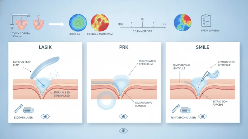
Clinical selection often balances corneal thickness, topographic regularity, and the magnitude of refractive error. For example, thin corneas or irregular topography may steer clinicians toward surface ablation or alternative treatments, whereas stable, moderate myopia with sufficient thickness might be treated with flap-based approaches or lenticule extraction. The presence of significant higher-order aberrations or prior ocular surgery can also guide the choice of procedure and the use of customized ablation protocols that aim to address optical irregularities beyond simple sphere and cylinder correction.
Healing responses vary among techniques, and recovery trajectories may differ accordingly. Surface procedures such as PRK typically require epithelial regeneration and may produce greater short-term discomfort and slower visual stabilization, often over days to weeks. Flap-based LASIK often yields faster visual stabilization in the early postoperative period for many patients, though both approaches can show ongoing refractive shifts for several months. SMILE recovery profiles may fall between these approaches for some patients, with reports indicating variable timelines depending on individual healing.
When comparing techniques, it is useful to view outcomes as probabilistic rather than guaranteed. Studies and clinical reports often summarize typical ranges of reduction in refractive error and rates of patient satisfaction, but individual results depend on preoperative measurements, surgical precision, and healing. Patients and clinicians commonly consider trade-offs among recovery speed, surface disruption, and the potential need for later enhancement procedures when selecting an approach. Further sections address technologies and planning that inform these choices.
Laser platforms used in refractive care often combine femtosecond and excimer technologies, each with distinct roles. Femtosecond lasers typically create corneal flaps or intrastromal cuts with high spatial precision, while excimer lasers ablate corneal tissue through a photochemical process that removes microscopic layers to reshape curvature. Treatment planning integrates refractive measurements, corneal imaging, and optical aberration data to define an ablation profile that targets intended changes in refractive power while seeking to preserve structural integrity.
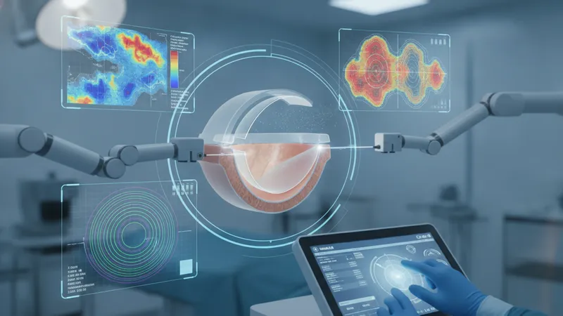
Diagnostic tools such as corneal tomography, topography, and wavefront aberrometry provide complementary information. Tomography yields elevation and thickness maps that help assess structural suitability and risk factors like ectasia; topography describes surface curvature patterns important for mapping astigmatism and irregularities; aberrometry quantifies higher-order aberrations that can affect night vision quality. Many clinicians use multiple modalities to triangulate a consistent surgical plan rather than relying on a single measurement.
Customization options aim to address individual optical imperfections. Wavefront-guided treatments use whole-eye optical measurements to tailor ablation to measured aberrations, while topography-guided approaches focus on surface irregularities by altering topo-driven contour. The choice among these guidance strategies may be informed by diagnostic findings; for example, irregular corneal surfaces may prompt topography guidance. Energy settings, ablation zone diameter, and intended correction depth are adjusted within safety margins derived from corneal thickness and biomechanical considerations.
Planning also considers enhancement contingencies and long-term stability. Clinicians typically document preoperative measurements and counsel that refractive outcomes can change over months to years, with a small fraction of eyes potentially requiring supplemental treatment. Imaging quality, repeatability of measurements, and agreement among instruments are practical considerations; inconsistent data often prompt further testing before finalizing a treatment plan. This iterative planning process helps set realistic expectations for outcomes and follow-up needs.
Eligibility assessment begins with a comprehensive ophthalmic history and objective testing. Stable refractive error over a period—often documented over 12 months—is typically considered an indicator of refractive stability, although acceptable intervals can vary by clinician. Key diagnostic tests include manifest and cycloplegic refraction, corneal topography/tomography, pachymetry for corneal thickness, and slit-lamp examination to assess ocular surface and lens clarity. These data help identify contraindications such as progressive corneal ectatic disease.
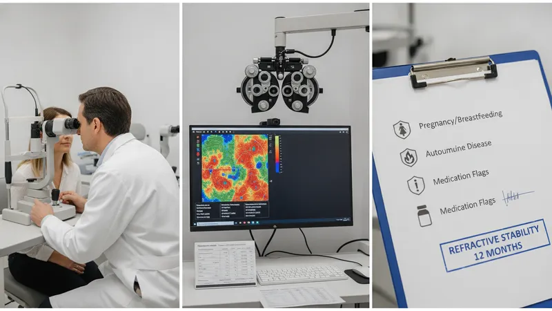
Systemic and ocular health factors may influence candidacy. Conditions that affect wound healing, such as certain autoimmune diseases, or medications that alter healing responses, may be relevant. Pregnancy and breastfeeding commonly prompt deferral of elective refractive procedures due to hormonal changes that can affect refractive stability. Age considerations typically aim to ensure ocular maturity and, in older patients, to evaluate lens status given the potential coexistence of presbyopia or cataract that may influence overall visual outcomes.
Tear film and ocular surface health are frequently assessed because postoperative dry eye symptoms can be a common transient effect. Tests may include osmolarity, tear breakup time, and assessment of meibomian gland function. If significant surface disease is present, clinicians often address these issues prior to proceeding, as optimizing the tear film can improve diagnostic measurement reliability and may influence postoperative comfort and visual quality. Patient lifestyle factors, such as occupational visual demands, are also part of the discussion as considerations rather than directives.
Informed consent and expectation management are components of pre-procedure care. Discussions typically cover the range of likely visual changes, potential transient effects like glare and fluctuations, and the possibility of needing enhancements. Measurements are often repeated to confirm stability, and alternative approaches may be considered if diagnostic data suggest elevated risk. These preparatory steps aim to align clinical decisions with an individual’s ocular characteristics and objectives while acknowledging that outcomes have inherent variability.
Recovery trajectories differ by technique but often follow a predictable early course. Many patients notice visual improvement within hours to days after flap-based procedures, while surface ablation may require several days to weeks for epithelial healing and visual stabilization. Over weeks to months, vision can continue to refine as corneal remodeling and neural adaptation occur. Follow-up visits at intervals such as one day, one week, one month, three months, and six months are commonly used to monitor healing and refractive stability, with exact schedules varying by provider.
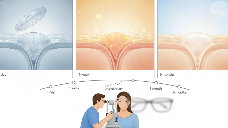
Expected outcomes generally aim to reduce dependence on corrective lenses, and many patients experience meaningful improvements in unaided acuity. Quantitative results vary based on initial refractive error, corneal anatomy, and healing. Some eyes may experience regression toward prior refractive states or residual refractive error that could be addressed with enhancement procedures under appropriate circumstances. Visual quality considerations—such as contrast sensitivity and higher-order aberrations—may also evolve during recovery and are part of outcome assessments.
Potential risks include transient symptoms such as dry eye, glare, halos, and fluctuations in vision. Less common complications can include infection, significant epithelial defects in surface treatments, or corneal structural weakening leading to progressive ectasia in susceptible individuals. Risk mitigation strategies often focus on thorough preoperative screening, conservative planning relative to corneal thickness, and postoperative monitoring to identify and manage issues early. Discussions about risks are typically framed in probabilistic terms rather than as absolute predictions.
Long-term considerations include the possibility of age-related changes such as presbyopia or cataract development that can affect uncorrected vision years after a refractive procedure. Patients and clinicians often consider how refractive surgery fits into an individual’s broader ocular health trajectory, including future needs for reading correction or cataract surgery. Ongoing follow-up and periodic eye examinations help to contextualize long-term vision changes and support decisions about any future interventions.